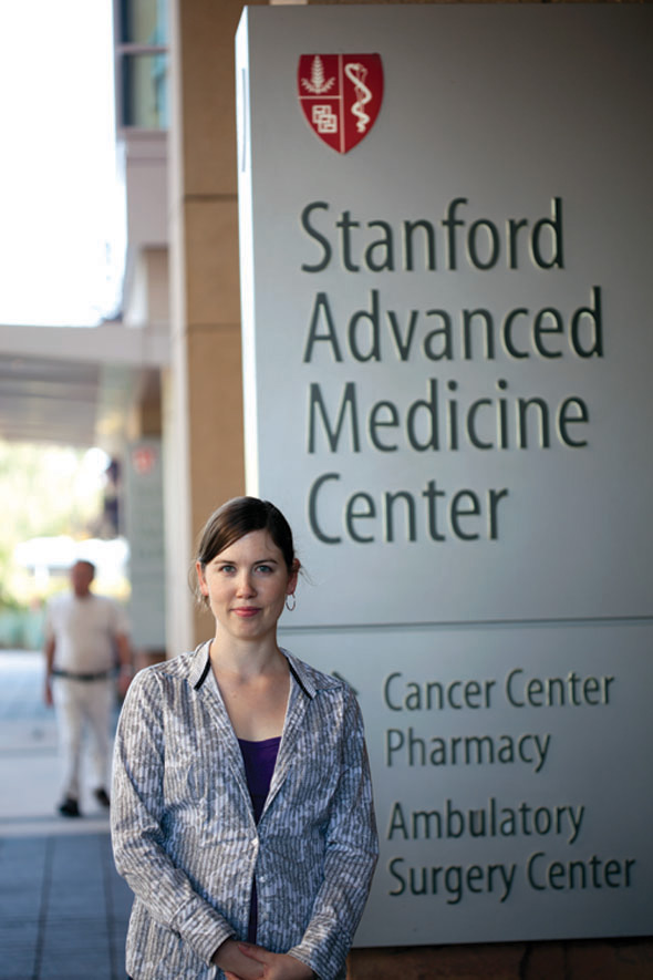
A physicist in the cancer lab
Nicole Ackerman thought she would always be a particle physicistuntil a newfound interest in biology drew her toward medical imaging. Her research on Cherenkov radiation, the blue glow from charged particles outracing light, could aid development of cancer treatments.
By Roberta Kwok
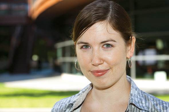 |
| Photography by Bradley Plummer |
Nicole Ackerman is a serious physics geek. As a graduate student at SLAC National Accelerator Laboratory, she ran computer simulations for EXO, the Enriched Xenon Observatory, an investigation into the nature of neutrinos; blogged for the particle physics website Quantum Diaries; and led lab tours. Her Gmail handle is neutrinoless, for the type of radioactive decay EXO is intended to detect. A tattoo of an equation called the Taylor series expansion circles her left arm because, she says, its the most beautiful thing Ive ever learned.
So what is she doing at the Stanford University School of Medicine? One of two researchers with particle physics training on an interdisciplinary team, Ackerman is helping to develop a new form of imaging that could be useful in testing cancer treatments.
In one sense, she hasnt strayed far from her roots: she studies a phenomenon called Cherenkov radiation, bluish light associated with fast-moving charged particles. Ackerman and other researchers are exploring whether Cherenkov light from radioactively tagged molecules could aid tumor imaging and drug development.
While her transition to biology has posed some challenges, Ackerman believes her training in particle physics simulations will allow her to make unique contributions to the field: It feels like its filling a niche that was previously empty.
Thinking outside the box
Ackerman became interested in physics in middle school, reading popular science books about quantum mechanics and string theory. As an undergraduate at the Massachusetts Institute of Technology, she traveled to CERN, the European particle physics laboratory near Geneva, to work on one of the detectors at the Large Hadron Collider, the most powerful particle collider in the world. Then she spent a summer at SLAC working on BaBar, an experiment investigating the universes puzzling shortage of antimatter, before starting her graduate studies there in 2007.
But after a few years, she began to wonder if she was suited to research in a field where a single experiment may span decades and involve hundreds or even thousands of people. To imagine spending my entire life on one projectI dont think I could do that, she says.
Then a new opportunity appeared.
At the June 2010 Meeting of Nobel Laureates in Lindau, Germany, Ackerman heard a speaker describe antibiotics binding to a molecule on a bacterium. The depth and complexity of molecular biology blew my mind, she says.
As she saw more talks and spoke to more biologists, for the first time in my life, I appreciated what biomedical research looked like, and how interesting it was.
About a week later, at a conference in Italy, she heard another talk about applications of particle physics in medical imaging and nuclear reactor monitoring. Ackerman had heard about these applications, but hadnt realized the work was happening in universities, not just in industry. She decided to switch fields and pursue applied particle physics for her PhD.
To learn more, Ackerman contacted scientists working in medical physics at the Stanford University School of Medicine, including molecular imaging researcher Ted Graves. When Graves told her his lab was studying Cherenkov radiation, she recalls, My reaction was to kind of squeak and say Cherenkov! Cherenkovs my favorite! She joined Graves lab and began working on a PhD project in fall 2010.
That strange blue glow
Cherenkov radiation is a really bizarre phenomenon, Ackerman says. Named for Russian physicist Pavel Cherenkov, who studied it in the 1930s, it occurs when a charged particle zips through a particular medium faster than light does. People get upset and say, well, things cant travel faster than the speed of light, she says. While thats true in a vacuum, light becomes more sluggish in materials such as water, and in some cases a charged particle can outrun it.
As the particle moves through the material, it distorts surrounding atoms by pulling or pushing away their electrons. The atoms relax back into place after the particle passes, releasing a bit of electromagnetic radiation. Normally, these emissions arent visible, just as tiny ripples on the ocean cant be seen from a plane. But if the particle breaks the light barrier, the radiation adds up to a bluish glow, analogous to a large ocean wave that is visible from far above.
To physicists, Cherenkov radiation is nothing new. BaBar, the experiment at SLAC, used Cherenkov light detection to help identify particles, and the South Pole observatory IceCube looks for Cherenkov light from particles produced by neutrinos hitting atoms in the ice. But in the medical community, it was largely ignored.
Eye-opening experiment
That changed in 2009, when a team led by researchers at Millennium: The Takeda Oncology Company in Cambridge, Massachusetts reported they could detect visible light emanating from mice injected with molecules labeled with a radioactive isotope. Radioactive labels are often used in medical research to follow molecules of interesta potential drug, for examplethrough an animals body, and in the clinic to evaluate cancer patients. Doctors may inject a patient with a radioactive form of glucose, which accumulates in sugar-hungry cancer cells and makes them stand out in an image.
My reaction was to kind of squeak and say Cherenkov! Cherenkovs my favorite!
 |
 |
 |
 |
I can look at the physics side of it and say, guys, this is your best-case scenario.
In this case, the team suggested the light was Cherenkov radiation, produced when the radioactive isotope decayed and emitted high-energy charged particles called positrons. The study suggested that the light offered a way to see radioactive decays in an animals body. The team called the new technique Cherenkov luminescence imaging, or CLI.
While researchers already obtain similar information with other techniques, such as positron emission tomography, or PET, CLI is cheaper. It provides a bridge between the world of PET and other nuclear imaging techniques and the world of optical imaging, says medical physicist Simon Cherry at the University of California, Davis. Some researchers hope to use CLI to image tumors in human patients during surgery, so doctors can check whether all the cancerous tissue has been removed before closing up a patient. However, this will be challenging since the light is very weak and cant penetrate far through tissue.
Running the numbers
Many CLI studies have been performed with mice. But Ackerman focused on studying Cherenkov light in the virtual world by running simulations with Geant4, software created primarily for particle physics. Developed in the 1990s, Geant4 allows researchers to simulate the behavior of particles. As a virtual particle moves through matter, the program essentially rolls the dice to determine what might happen at a given instant, repeats the process to plot the particles path and decay, and then does this for many particles. Geant4 has found a variety of uses outside particle physics, such as estimating the amount of radiation astronauts receive in space and verifying radiation treatment plans for cancer patients.
Ackerman had already used Geant4 during her time on EXO to simulate parts of the data-gathering process. In Graves lab, she focused on radioactive isotopes called alpha emitters, which scientists hope to use to kill cancer cells but currently dont have a good way of imaging. With her simulations, she was able to confirm that the alpha emitter actinium-225 could also indirectly produce Cherenkov light through high-energy electrons released by its daughter isotopes. Scientists developing alpha-emitter treatments could use Cherenkov imaging to watch a drugs path in mice and ensure its reaching the tumor, she says.
Ackerman has also used Geant4 to study other potential cancer treatments. For example, scientists hope to increase the effectiveness of radiation treatments by injecting patients with gold nanoparticles that accumulate in the tumor and intensify the radiation dose to the area. Ackermans simulations allowed her to estimate the size of this effect: It could increase the radiation dose to the tumor cells nuclei by about 2.5 times. By trying different nanoparticles in Geant4 and determining an upper limit on the effect, Ackerman might be able to save researchers time and money in the lab.
I can look at the physics side of it and say, guys, this is your best-case scenario, she says. If youve gotten that, you dont need to keep trying to make different types of nanoparticles to see if you do better.
Geant4 isnt the perfect tool for Ackermans research. The standard Geant4 models of physics processes typically deal with higher energies than those seen in radiation treatments for cancer. But Ackerman plans to investigate how much these limitations affect her data and whether the models could somehow be modified to address them. She also hopes to program a virtual mouse into Geant4 so she can simulate processes in mice, the standard lab animals.
A new start
According to Graves, the traditional curse of biology is that its a qualitative science; physicists like Ackerman bring a more quantitative mindset to the research. But Ackerman has also been eager to learn about biology. Thats refreshing and important, says Rehan Ali, a postdoc in Graves lab. If youre coming from a particle physics background and want to contribute to biomedicine, you have to really spend time getting to know the field first, and shes done that.
Making the transition from particle physics to biology hasnt been entirely easy. After signing up for an imaging anatomy class that required her to identify organs, Ackerman says, I thought, oh crap, how am I ever going to learn all our squishy bits inside? Because they all kind of look the same. And when trying to simulate the behavior of an imaging instrument, she ran into difficulties getting information from the manufacturer. Coming from particle physics, the guy next to you wrote the code and the guy on the other side built the system, she says. So its been very strange trying to model this system and not knowing all the details.
Even in her new environment, Ackerman maintains ties to her particle physics past and follows news in the field. While she misses the particle physics community, she says she likes contributing to a field where her simulation skills are less common. I might be the only one thinking about some of these details, she says. If her alpha-emitter studies help researchers develop a cancer treatment more quickly, thatll make a difference to people I know, she says. The work I do matters more.
Click here to download the pdf version of this article.