The same particle-physics technology used to understand the universe is also used to improve health and medicine. Accelerators and detectors play an important role in diagnosing disease, shrinking tumors and sterilizing medical equipment. Large-scale computing makes it possible to determine which potential new drugs are most likely to work before starting large-scale human trials. And particle-physics-trained scientists serve as medical physicists, making sure it all works as planned.
Sterilizing instruments and supplies
Particle physics technology can be used to disinfect syringes, bandages, scalpels, stethoscopes and other tools without damaging them. Medical equipment is sent through a series of small particle accelerators and bombarded with beams of electrons or X-rays. In a matter of seconds, the beams eradicate any surface microbes.

Distributed and grid computing
The World Wide Web is not the only computing advancement to come out of particle physics. In order to cope with the huge amount of data produced by experiments, particle physicists developed a network of grids allowing multiple users to share computing power and storage capacity. The grid concept has a number of uses in the medical field, including screening drug candidates to determine which ones are most likely to fight disease.

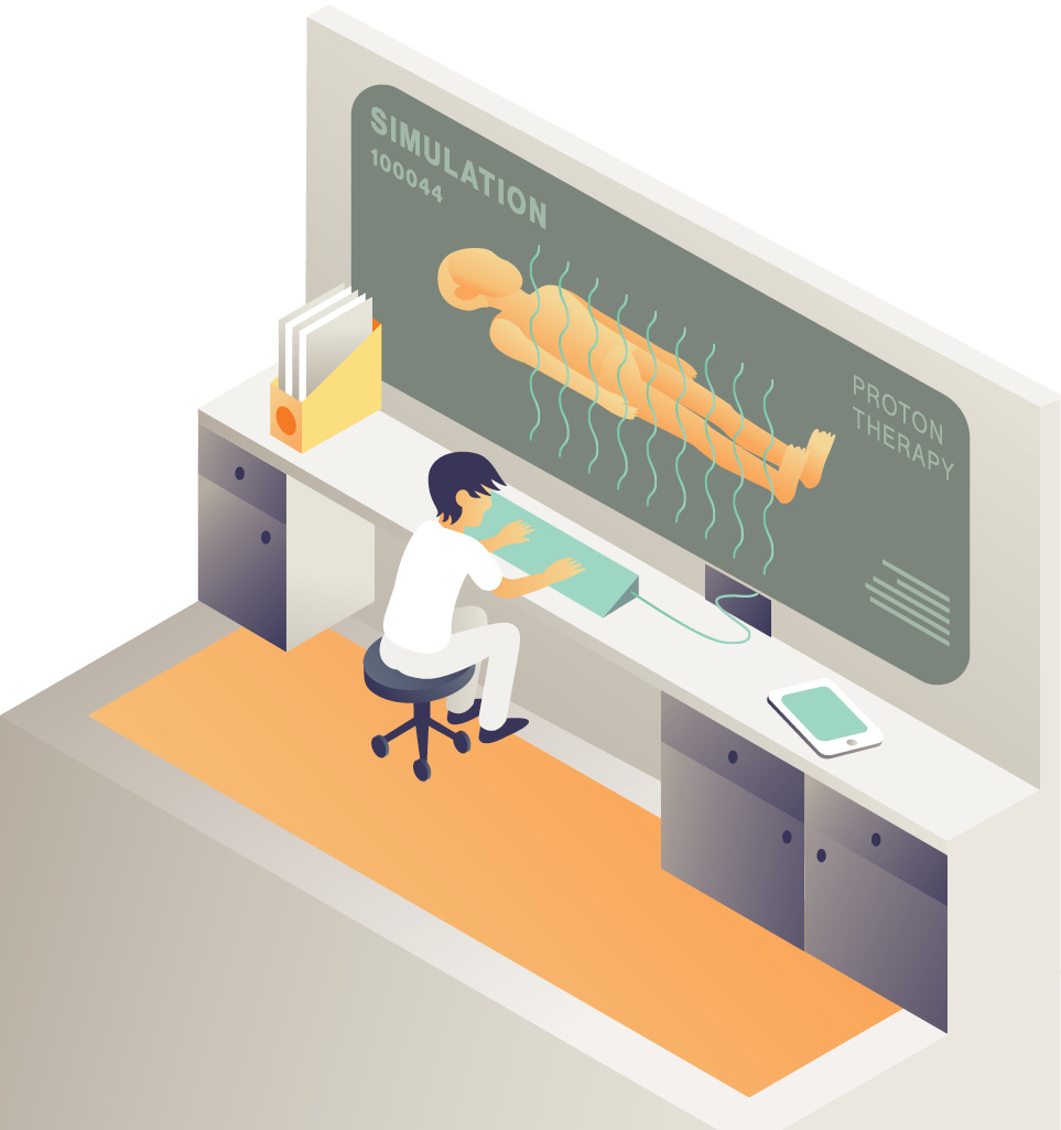
Simulation
Practice makes perfect, and when it comes to our health, the closer to perfect, the better. So some doctors and medical physicists are designing treatment plans using modeling tools developed for particle physics to predetermine the electromagnetic and nuclear interactions of particles with tissue. In radiation therapy, this software can help doctors understand what will happen when a beam of particles passes through a patient’s body.
Semiconductors
In the heart of particle physics detectors around the world, hundreds of detectors made with silicon semiconductors splay out around particle collision points, tracking charged particles to create pictures of their paths. Physicians make use of this semiconductor technology in many medical devices, including semiconductor lasers. These discrete beams of high-intensity light are perfect for delicate operations like eye surgery.
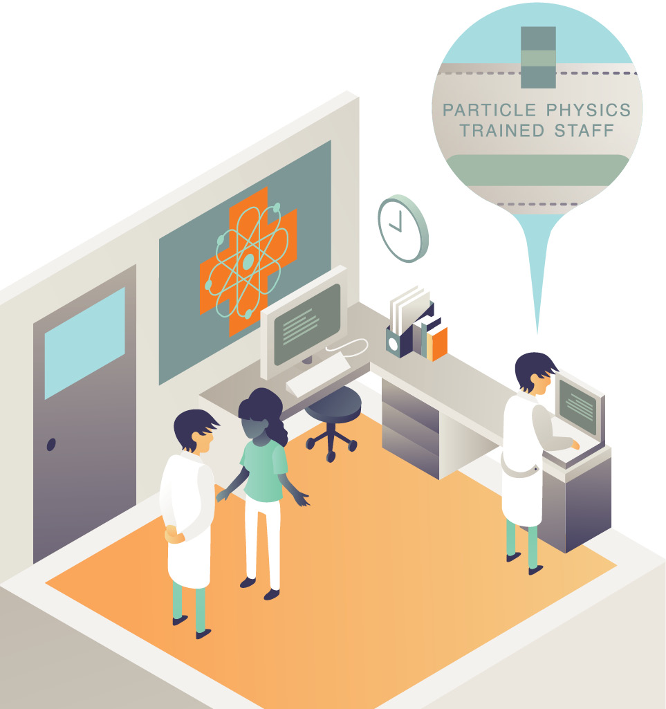
Particle-physics-trained staff
Many particle physicists can be found inside hospitals and clinics. Particle physicists who cross over into the medical field often come with extensive training in the operation and maintenance of accelerators. With their thorough understanding of particle beams, these scientists are highly valued as specialists who manage the medical imaging systems that detect tumors and who operate the accelerator beams that kill cancer cells.
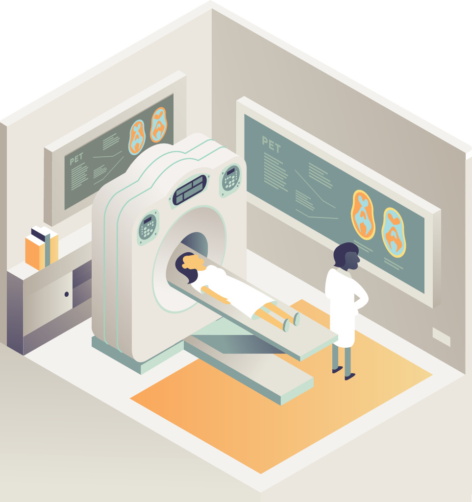
PET
PET scanners are common tools that let medical professionals examine organs and tissues inside the body. The PET scanner’s genealogy traces back to detector technologies developed in the 1980s to identify individual photons in particle physics experiments. It may sound strange, but PET scanners use antimatter produced inside the body. When a special tracer is injected into a patient, a type of radioactive decay occurs, emitting positrons—the antimatter counterparts to electrons. These positrons annihilate with nearby electrons, releasing bursts of photons. The photons are detected and compiled into three-dimensional images.
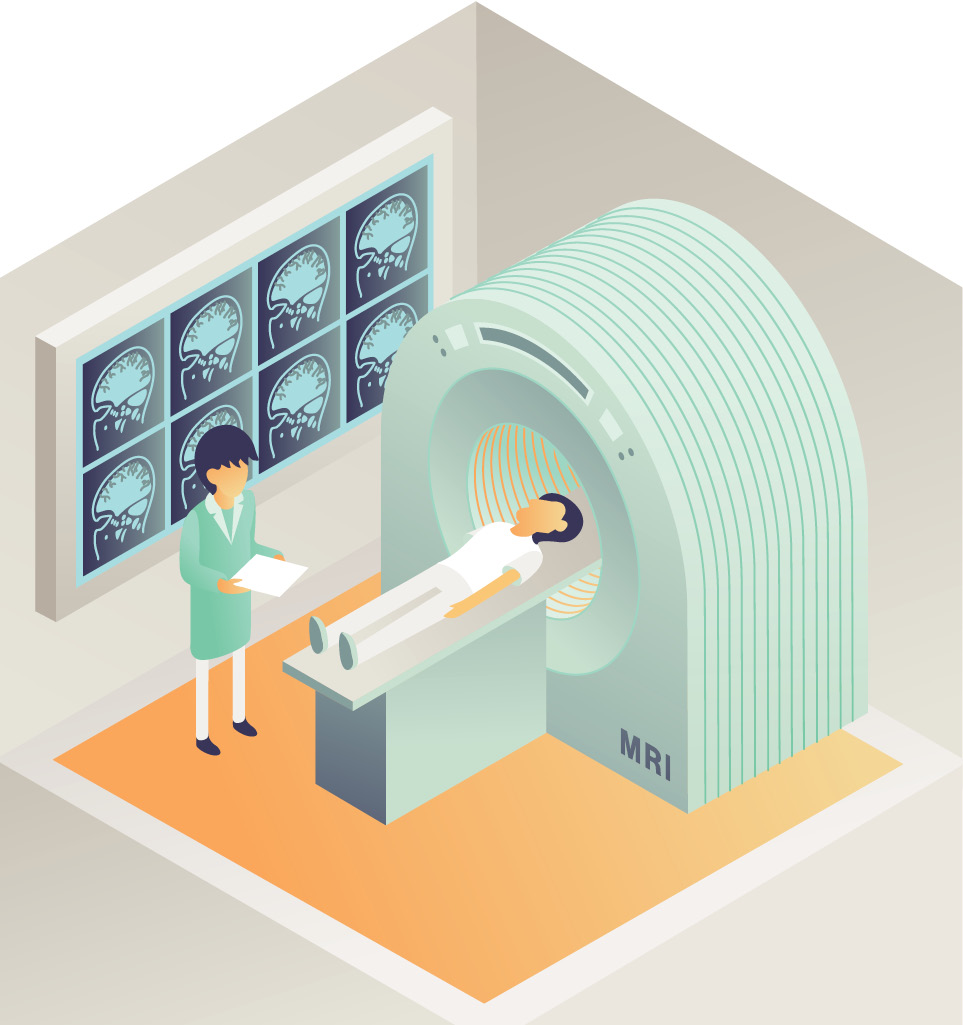
MRI
Magnetic resonance imaging, the basic principles of which emerged from early research in physics, is more discerning than traditional screening, which sometimes can’t make out tumors hidden within dense tissue. When a patient is subjected to the powerful magnetic field inside an MRI machine, atoms inside his body line up in the direction of the field. A radio frequency current is temporarily switched on, causing the protons inside those atoms to flip around until the radio frequency is removed. At that point, the protons pivot back into place—each at a different rate. The varying rates are measured, allowing scientists to determine what’s happening inside the living tissue.
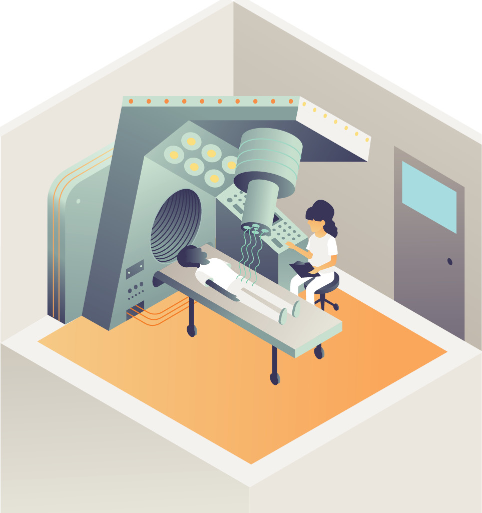
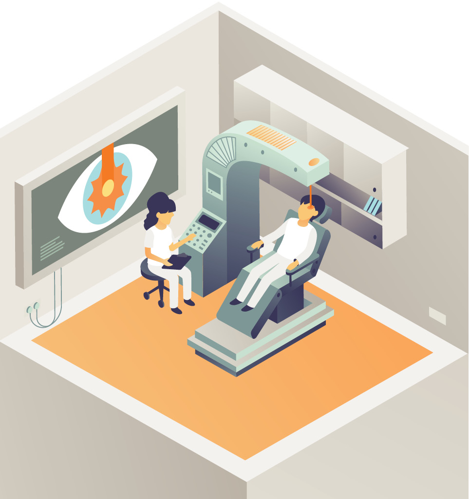
Cancer treatment
One of the most effective techniques to fight cancer uses the same technology particle physicists employ to accelerate particle beams to nearly the speed of light. There are more than 17,000 particle accelerators worldwide used for the diagnosis and treatment of disease. Doctors exchange a scalpel for a beam of charged particles, which they aim at cancerous tissue, killing malignant cells by destroying DNA strands in the nuclei while sparing the surrounding healthy tissue.